NOTE: for identification of specific species, there are links
at the bottom of the "growth rings" page (LINK) that will
take you on a guided tour of detailed images for about 450 species
BUT ... it is helpful to know the basics of parenchyma before you go there
PARENCHYMA
Parenchyma cells are wood tissue that is typically described as brick shaped or roughly football shaped (but with flat surfaces instead of smooth rounded ones) in form and which are used by the tree to store and distribute carbohydrates (tree food). These are sometimes more elongated than is indicated by the descriptions "brick shaped" and "football shaped", but what is more important to keep in mind is that these are MICRO descriptions and are about characteristics that are never visible to the naked eye and often not even with a 10X loupe. This article is about parenchyma characteristics that ARE visible with a 10X loupe.
Parenchyma cells have two fundamental types, axial and ray. Axial cells run up and down the length of the tree and ray cells run radially. Rays are very important in wood identification but all discussion of parenchyma cells in this section is of axial cells, not ray cells.
Axial parenchyma cells are of two fundamental types. They can occur in various forms where they are touching pores, in which case they are called paratracheal or they can occur away from pores in which case they are called apotracheal. "Tracheal" means tube-like, which is a good description of wood pores and para and apo are Greek prefixes meaning "next to" and "away from", so these names make good sense once you see the derivation. The terms apotracheal and paratracheal are used throughout this section, so you will find it helpful to remember them. There are a ton of terms that are used to describe various groupings of parenchyma cells and there is overlap among some of the terms. That is, a given species might well have 2 or even 3 types of parenchyma cell groupings. In such cases there is usually a clearly predominant trait that dictates which term is used as the primary descriptor for the species. I have pointed out several such combinations in the illustrations in this section.
NOTE: I have found numerous contradictory/conflicting definitions and examples of parenchyma types on the Internet, even among university sites and other apparently knowledgeable sites. Where possible, I have followed the definitions given in Identifying Wood by Bruce Hoadley
The various forms of parenchyma cells include (NOTE: the descriptions following the "---" are generally based on end grain cross sections)
apotracheal --- an adjective meaning "away from the pores" (contrast to paratracheal)
aliform --- wing-like paratracheal groups of cells that can look like an airplane or a lozenge in end grain cross section
axial --- the length goes up and down the tree (same as longitudinal)
banded --- full bands of cells that are most often apotracheal but may be paratracheal
boundary --- the same as marginal
confluent --- paratracheal cells connecting two to several pores
diffuse --- apotracheal and widely spaced, generally not visible with a 10X loupe
diffuse-in-aggregates --- apotracheal and widely space but in groups that might be visible with a 10X loupe
epithelial --- surrounding resin canals in softwoods
fusiform --- longitudinal but without cell cross-walls
initial --- marginal, at the start of the earlywood
longitudinal --- the length goes up and down the tree (same as axial)
vasicentric --- parenchyma cells form a complete circle around / touching the pores
wing shaped aliform --- wing-like paratracheal groups of cells that look like an airplane
zonate --- confluent parenchyma that is particularly solid and well formed (this seems to be an ill-defined term and I have seen conflicting definitions and examples)
apotracheal parenchyma --- "apo" means "away from" and "tracheal" mean "tube-like", so this is a very descriptive name for parenchyma cells that stand apart from the pores in wood. There are three basic forms that this can take:
banded --- full bands of cells that go all the way around the tree, like growth rings, but more frequent
diffuse --- cells that are here and there in no distinct pattern and that are generally too small to see with a 10X loupe
diffuse-in-aggregates --- cells that are here and there in small groups, usually in a band but otherwise in no distinct pattern and that are sometimes too small to see with a 10X loupe
marginal --- starting the earlywood (initial) or ending the latewood (terminal) and may be close to pores but are not directly associated with them
Note that "banded" is actually much more complicated than the simple statement above. First, banded may NOT be continuous all the way around the tree but rather may be briefly broken in places. Second, banded is only USUALLY, not exclusively, apotracheal; there are common examples where banded groups are paratracheal.
Apotracheal is to be contrasted with paratracheal which means "next to the pores".
aliform parenchyma --- "Aliform" means "wing-like", so not too surprisingly this is an arrangement of parenchyma cells that, when seen in transverse cross section, form either (1) thin wing-like lines attached to the sides of pores or (2) clumps of cells that taper off as they move away from the pores. The first of these constructs looks a bit like a WWII fighter plane and is called "winged aliform parenchyma" and the second is somewhat football-shaped but is formally called "lozenge shaped aliform parenchyma".
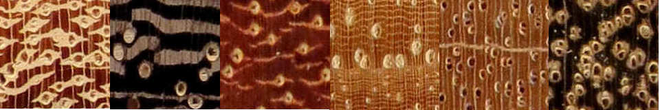
The first pic is winged aliform parenchyma in a 1/4" square end grain cross section of ramen (Gonystylus spp.) shown here at 12X. The color was slightly over-saturated to make the parenchyma lines (the airplane wings) stand out more clearly.
The second pic is lozenge shaped aliform parenchyma in a 1/4" square end grain cross section of kempas (Koompassia malaccensis in this case) shown here at 12X. This is a weak lozenge shape.
The third pic is lozenge shaped aliform parenchyma in a 1/4" square end grain cross section of kempas (Koompassia malaccensis in this case) shown here at 12X. This is a strong lozenge shape.
The fourth pic is lozenge shaped aliform parenchyma in a 1/4" square end grain cross section of ramin (Intsia spp.) shown here at 12X. This is a strong lozenge shape.
The fifth pic is winged aliform parenchyma in a 1/4" square end grain cross section of padauk, right at the beginning fo the sapwood shown here at 12X.
The sixth pic is winged aliform parenchyma in a 1/4" x 1/6" end grain cross section of narra (Pterocarpus indicus) shown here at 12X
The seventh and last pic is lozenge shaped aliform parenchyma, extending in a couple of pores to winged aliform parenchyma, in a 1/4" x 1/6" end grain cross section. There is no confulence in this piece so I didn't do red or blue circles. Note also the marginal parenchyma lines
In each of these pics, I have used a blue circle to show an example of a solitary aliform group and a red circle to show where these have merged or extended into each other to become confluent (which doesn't ALWAYS happen, as is shown in the last pic where the bubinga shows no confluence)
Although, as you can see, some of the individual lozenge shaped cell groups are almost circular and so might be thought to be vasicentric groups; the difference is that these aliform groups form much thicker rings around the pores than do vasicentric groups.
banded parenchyma --- Bands of parenchyma cells running all the way around a tree, having the same orientation as the growth rings but there tend to be many of the "bands" within a given growth ring.
Banded parenchyma in mockernut hickory, sapele, ekki, and cocobolo with all of them presented in 1/4" x 1/6" end grain cross sections shown here at 12X. The ekki was not subject to my cleanup so shows saw marks. As you can see, in practice the "bands" are not always exactly circular but rather are a bit erratic in their orientation over short distances, making up a rough band over longer distances.
Although banded parenchyma is primarily categorized as apotracheal that is because they don't HAVE to be next to pores, but as you can see in these examples, they are often paratracheal.
Sometimes the appearance of the bands is not totally continuous. Whether this is because of an actual break in the lines of cells or just a thinning that makes them hard to see with a 10X loupe, I don't know for sure but my belief is that it is a real occurance, not just an apparent one. For example, the blue circle in the first pic is around an area that appears to have a couple of breaks in the bands. There is another form called diffuse-in-aggregates that can take the form of broken bands very similar to banded lines except that they tend to be much more broken up and sparse.
confluent parenchyma --- Confluent literally means "flowing together or merging" so not surprisingly that's just what it means with parenchyma cells. Specifically, the parenchyma cells connect two more more pores that are themselves surrounded by parenchyma cells. The type of cell groupings that make up the banding can be various types, such as aliform and banded. Note that there are woods (particularly elm and hackberry) that have wavy bands of pores that look like thick groups of confluent parenchyma but are not.
Confluent parenchyma examples in 1/4" x 1/6" end grain cross sections show here at 12X.
American elm / Ulmus americana
wenge / Millettia laurentii
wawabima / Sterculia rhinopetala
sen / Kalopanax septemlobus
white ash / Fraxinus americana
honey locust / Gleditsia triacanthos
purpleheart / Peltogyne lecointei
merbau / Intsia bijuga
All the woods in the first row have very heavy, obvious confluence. The second row is examples of woods that have lesser amounts of confluence, with each pic having a single instance of confluence circled in blue and an instance of vasicentric parenchyma circled in green.
In the purpleheart, a red circle shows a single construct which is clearly aliform parenchyma of the lozenge type and a green circle shows one that is aliform but if viewed out of context the green-circled pore might well be considered vasicentric. The white arrows point to marginal parenchyma. This is just another example of how woods have more than one type of parenchyma.
diffuse parenchyma --- Diffuse individual parenchyma cells that are too small to see with a 10X loupe and thus are of no interest in this discussion.
diffuse-in-aggregates parenchyma
This section had incorrect information and is being updated
--- Diffuse groups of apotracheal parenchyma that are sometimes big enough to see with a 10X loupe. Sometimes these take the form of what appears to be banded parenchyma except that the bands are clearly broken up and tend to be more sparse than full banded groups (which completely ring the tree).
diffuse-in-aggregates parenchyma (the thin horizontal lines) in a set of 1/4" x 1/6" end grain cross sections of shown here at 12X. The woods are claro walnut, butternut, machiche, and English brown oak. Had I just looked at the butternut or the walnut end grain without benefit of any research, I would likely have thought they were banded, but several sources including Hoadley say that Juglans parenchyma is diffuse-in-aggregates, showing constructs that are exactly like what is in these pics except that their pics are better than mine and tend to show the breaks more clearly. Personally, I consider this description to be misleading. The DEFINITIONs of diffuse-in-aggregates include such terms as "diffuse" and "widely spaced" but the bands do not seem diffuse or widely spaced and in fact look to me exactly like banded parenchyma. In the English brown oak, on the other hand, the spacing is wider. In the machiche there is relatively little diffuse-in-aggregates parenchyma and I've put in a pointer to two of them.
diffuse-in-aggregates parenchyma in a pair of 1/4 square end grain cross sections of padauk shown here at 12X and circled in green. To once again show the overlap among types, I have also, on each of these pics, circled wing-like aliform parenchyma in red and confluent parenchyma in blue. The "widely spaced" part of the definition clearly applies here since the GROUPS of parenchyma are widely spaced, but the "diffuse" here, as in the butternut above, eludes me. These are clearly NOT banded parenchyma but the whole definition of diffuse-in-aggregates seems hard to reconcile with the examples it's used on (and these are not arbitrary choices based on MY decisions, they are species used as illustrations in references such as Hoadley)
epithelial parenchyma --- Parenchyma cells that surround a resin canal in a softwood. Resin canals can be axial or radial and in both cases they are surrounded by epithelial cells. The resin canals themselves are tubular voids in the wood and the surrounding parenchyma cells bleed resin into them. I am not aware of any way these are useful in identifying wood with only a 10X loupe, as they are much smaller than the resin canals (which are themselves more useful in wood identification).
fusiform parenchyma --- longitudinal parenchyma cells sometimes have what are called "cross-walls" extensions from the end of one cell to the beginning of another cell. These are never visible with a 10X loupe so are of no interest in this discussion. Fusiform cells are the same as other longitudinal parenchyma except that they do not have the cross-walls
marginal parenchyma --- parenchyma cells that exist at the very beginning (initial parenchyma) or very end (terminal parenchyma) of a growth ring, sometimes in small amounts here and there, sometimes as solid lines right at the border between the latewood of one growth ring and the earlywood of the next ring and help distinguish that demarcation. Sometimes even the full bands are not visible even with a 10X lens but in some species they form a visible line, sometimes a solid line, more often a very rough, vague line) at the boundary between early and late growth I do not know how to determine whether such a line is initial or terminal. That is, a line of marginal parenchyma cells can be very distinct but as far as I personally know how to judge, such a row could belong to the end of a growth ring (late latewood) or the beginning of a growth ring (early earlywood). In any case, as far as wood identification is concerned, the distinction is irrelevant. The full lines of marginal parenchyma are very similar to banded parenchyma except there is only one band per growth ring and it's at the margin between latewood and earlywood. Also, they tend to be less erratic in direction than banded parenchyma.
marginal parenchyma lines in a 3/8" square end grain cross sections of two pieces of Honduran mahogany (Swietenia macrophylla) and then one of Cuban mahogany (Swietenia mahagoni) shown here at 8X. Often, the marginal parenchyma lines in the Swietenia species are extremely coherent and distinct as shown in the first pic but even when they are more diffuse/fuzzy, they remain coherent (as shown in the second pic), unlike those in African mahogany (as shown directly below) which can go from very distinct to totally indistinct or even non-existent in a short distance.
marginal parenchyma lines in a 3/8" square end grain cross section of African mahogany (Khaya spp.) shown here at 8X. Marginal parenchyma lines in African mahogany are sometimes as distinct as those in the Swietenia species, but more often they are more diffuse and vague, often changing from one form to the other in the same line. Notice the lower line in the first pic which is fairly strong on the right but trails off on the left side. Notice in particular how vague the lower line is in the second pic. I have seen some African mahogany where a marginal parenchyma line will be strong and distinct in one area and the same line one inch away will have become so totally vague as to merge into and become indistinguishable from, the pores as shown directly below.
a one-inch long end grain cross section of African mahogany, shown at about 9X, with a marginal parenchyma line showing how such a line can go from distinct (on the left) to indistinct or even non-existent (on the right) in a very short distance (when you look at it, please keep in mind that this IS only a 1" wide sample even though it is shown 9" wide here). If you were to look at just the right-most 1/3 of this pic, you would conclude that this wood has no parenchyma lines.
1/4" square end grain cross sections of tulip poplar (Liriodendron tulipifera), cucumber magnolia (Magnolia acuminata), and butternut, all shown at 12X, with marginal parenchyma lines. In these species, the lines can be hard to see with the naked eye, as in the poplar samples shown, or readily visible to the naked eye, as in the butternut sample but in all cases they are visible with a 10X loupe. For these species, growth ring boundaries are normally easily distinguished, even if sometimes a bit vague as in the poplar sample. Sometimes this is less because of the marginal parenchyma lines than the fact that there is a distinct darkening of the wood at the end of the latewood and a lightening at the beginning of the earlywood as seen in the tulip poplar.
A one inch long flat cut area of Honduras mahogany, shown at about 5X, with marginal parenchyma lines that are distinct enough to see with a 10X loupe and even without a lens if you know what you are looking at. Being able to see marginal parenchyma lines in a flat cut section is rare and the Swietenia species are among the few that sometimes exhibit this and it is one of the ways that the Swietenia species (Honduran and Cuban mahogany) can sometimes be distinguished from the Khaya species (African mahogany) which almost always have fuzzy marginal parenchyma lines, not sharply defined ones like those found in the Swietenia species.
Marginal parenchyma (circled in blue) specifically identified by Hoadley as terminal, shown in two 1/4" square end grain cross sections of Spanish cedar (Cedrela fissilis) and red mountain Spanish cedar (Cedrela montana) shown here at 12X. Also shown circled in green are instances of vasicentric parenchyma. Personally, I would have said these have to be initial parenchyma, not terminal, because interspersed among the parenchyma cells, on the first pic at least, are large earlywood pores. Possibly the parenchyma cells are not the entire light-colored area but just a thin row at the bottom of that area, in which case it then becomes reasonable that these could be terminal, not initial. At any rate, Hoadley knows more about this stuff than I do.
Marginal parenchyma (circled in blue) specifically identified by Hoadley as terminal, shown in two 1/4" square end grain cross sections of Ceylon satinwood (Choloroxylon swietnia) shown here at 12X. Clearly, these are barely visible with a 10X loupe.
paratracheal parenchyma --- "para" means "next to" and tracheal means "tube-like", so this is a very descriptive name for parenchyma cells that are close to pores in wood. There are three basic types that this form can take:
aliform --- wing-like paratracheal groups of cells that are clustered around pores and extend out from pores. These can look like an airplane or a lozenge in end grain cross section
confluent --- paratracheal cells connecting two to several pores. These can range from skinny lines to thick lines
scanty --- parenchyma cells that are next to pores but very few in number (thus the "scanty") and tiny (not visible with a 10X loupe)
vasicentric --- "parenchyma cells that form a complete circle around / touching the pores but that, unlike aliform groups, do not extend away from the pores
Paratracheal is to be contrasted with apotracheal which means "away from the pores".
reticulate parenchyma --- Banded lines of parenchyma that go all the way from one ray to the next and generally cross many rays, with the spacing and size of the parenchyma lines and the ray being about the same. This construct looks like a mesh or a fish net which makes good sense, since "reticulate" means "net-like".
The examples for this construct that I was able to find in my own wood pics were right at the limit of what you can see with a 10X loupe, so in both of these images, I have blown them up beyond the natural limit of the image and then sharpened them up a bit with some image manipulation. This was to make it very apparent right here what the construct looks like; you COULD see it in a 10X loupe without this manipulation but it would not have been as clear here as I wanted for this illustration.
Both of these pics are 1/8" end grain square cross sections that have been blown up to 24X (the original image was only good to 12X). The first one is hickory (Carya spp.) and the second is mockernut hickory (Carya tomentosa)
scalariform parenchyma --- When a wood has what would otherwise be called reticulate parenchyma but the rays are very fat compared to the lines of parenchyma cells and the horizontal lines are a bit curved, then the overall look is said to be a bit like a fish scales (thus the "scalariform"). Although I have not seen mention of it in formal reports, I have noticed in this form that the short parenchyma lines between rays always seem to bow inwards slightly (which is downwards in these pics since the pith is downwards) in all cases but the pore location relative to the parenchyma lines will vary. My expericence is that, as you can see in the samples shown directly below, in Australian lacewood, and ONLY in Australian lacewood, they tend to be directly in line with the parenchyma lines whereas all of the other woods that I have samples of they seem to hang off of the bottom of the parenchyma lines. I also note that unlike reticulate parenchyma lines, which are predominantly apotracheal and are categorized as such, these lines are reported as paratracheal at least as often as they are apotracheal and in my experience, as you can see in the samples shown below, they are always paratracheal since the where the pores are "hanging off of the parenchyma lines", the pores do touch the lines and of course in Australian lacewood the lines are directly bisected by the pores.
Scalariform parenchyma in 1/4" square end grain cross sections, presented here at 12X
"seemingly marginal" --- fairly often, woods will have what appears to be marginal parenchyma but is listed as "seemingly marginal", but which I take it to mean that the bands may not actually be on a growth ring boundary. I have not found this term to be widely used but that might be because other authorities know for sure whether or not the bands on a given species are marginal or not.
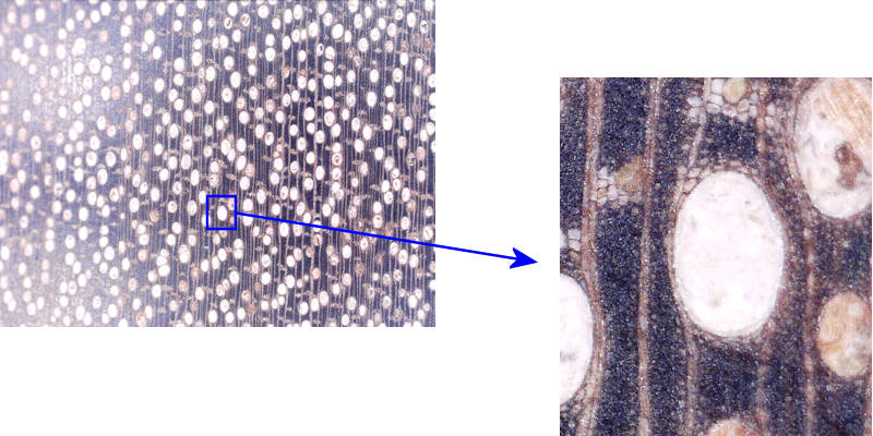
scanty parenchyma --- paratracheal parenchyma cells that are very few in number (thus the "scanty") and generally tiny. As these are too small to be seen with a 10X loupe, they are of no interest in this discusion. Because they are next to the pores, they are paratracheal. If they completely surround the pore, they are vasicentric.
Here's an example of scanty vasicentric parenchyma in a piece of meranti. At 10X (seen on the left), the small amount of parenchyma cells next to the pore (seen in detail on the right) are indistinguishable from the surrounding tissue.
vasicentric parenchyma --- "vasi" means "vessel" and one of the meanings of centric is "concentrated around", so this is a good descriptive term for parenchyma cells that are all around a pore, with the pore at the center. Vasicentric cells, when they exist around the pores in a given wood, range from very skinny to really fat.
Vasicentric parenchyma cells in 1/4" x 1/6" end grain cross sections, presented here at 12X, around the pores in idigbo and koa, moderately thick, and then particularly fat vasicentric parenchyma around the pores in kelobra and alibizia. An enlarged view of an area of the albizia sample on the right is shown below.
More detail on an area of fat vasicentric parenchyma on the same piece of albizia shown in the composite image directly above, showing the individual parenchyma cells around a pore. At 10X (on the left) it's easy to see the blob of parenchyma cells around the pore but the individual parenchyma cells shown at higher magnification (on the right) are not distinguishable at 10X. For several more such images, go here: vasicentric parenchyma examples.
.jpg)
.jpg)
.jpg)
.jpg)
.jpg)
.jpg)
.jpg)
.jpg)
.jpg)
.jpg)
.jpg)
.jpg)
.jpg)
.jpg)
.jpg)
.jpg)
.jpg)
.jpg)
.jpg)
.jpg)
.jpg)
.jpg)
.jpg)
.jpg)
.jpg)
.jpg)
.jpg)
.jpg)
.jpg)
.jpg)
.jpg)
.jpg)
.jpg)
.jpg)
.jpg)
.jpg)
.jpg)
.jpg)
.jpg)
.jpg)
.jpg)
.jpg)
.jpg)
.jpg)
.jpg)
.jpg)
.jpg)
.jpg)
.jpg)
.jpg)
.jpg)